Research News
Frequently, one wants to perform several analyses on a single piece of tissue in order to draw conclusions about the presence of certain proteins in combination with the function and anatomical properties of the cells. Neurobiologists, in particular, are often faced with this challenge: When they want to reconstruct the synaptic connectivity of neurons and likewise obtain functional information about neurons and synapses, 3D electron microscopy (EM) and immunohistochemistry (IHC) have to be combined. Until now, it has not been possible to combine both procedures in thick tissue samples. The limiting factor was mainly the reliable labeling of structures deep within the sample without harming the ultrastructure of the tissue. Researchers at the caesar institute now describe a protocol that avoids the need for tissue permeabilization in thick tissue sections, a major constraint to correlative pre-embedding IHC and EM. In their study, they nicely show proof-of-principle experiments combining two-photon imaging of protein distributions and 3D electron microscopy. The protocol they developed enables the correlation of function, cell type identity, neuronal morphology and synaptic connectivity within local circuits and can be certainly further developed for other tissue samples and organisms. Incorporating the molecular properties of neurons into reconstructions of whole brain neural connectivity will be the next important challenge. Further, this opens up deep analyses of human tissues, for example.
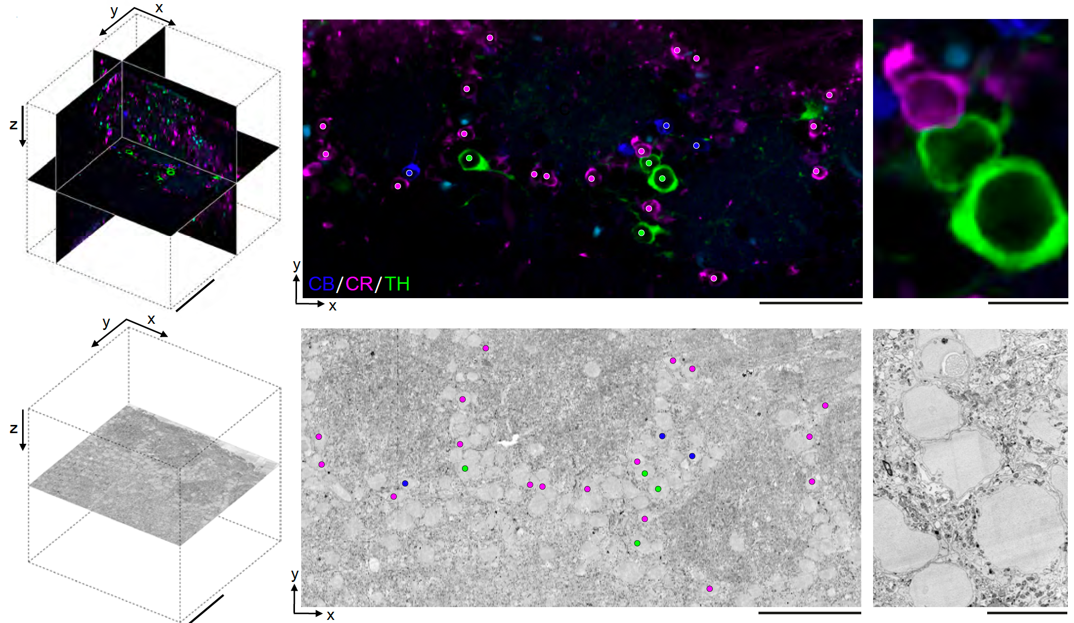
Correlative microscopy of various interneurons in the mouse olfactory bulb.
For further information please contact: